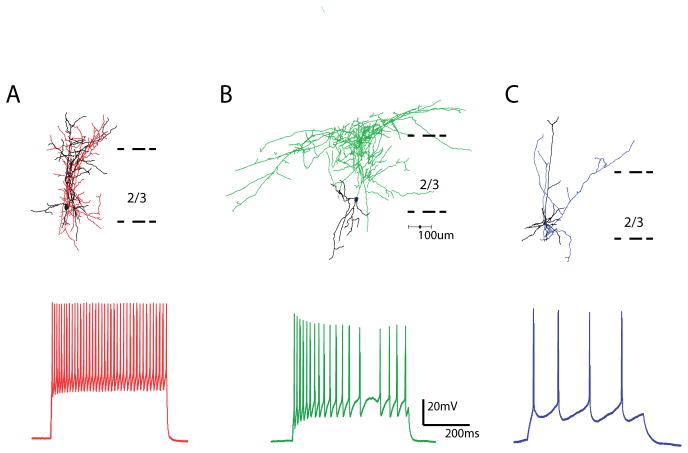
Figure 4. Morphological and physiological characterization of interneurons.
A, Reconstruction of a PV GFP positive basket cell (top), with typical fast spiking physiology (below). Axons in red, dendrites in black. B, Reconstruction of a SOM GFP positive martinotti cell, with typical accommodating firing pattern shown below. Axons in green, dendrites in black. C, Reconstruction of a layer 2/3 PC, with characteristic regular firing pattern shown below. Axons in blue, dendrites in black.

