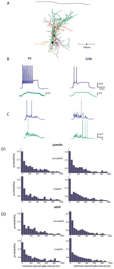
Figure 6. Electrical coupling does not influence synchrony of interneurons.
A, Anatomical reconstruction of 2 pvGFP cells patched within 60μm of one another that were gap junction coupled (axons of cell 1 are in green, dendrites in red, Axons cell 2 in blue, dendrites orange) B, Intracellular current injections in a PV neuron and a SOM neuron at 2x rheobase (blue traces top), with a nearby electrically coupled neuron patched within 100μm (green traces, bottom). C, Representative traces from a pair of PV interneurons, and SOM interneurons, respectively, in response to thalamic stimulation. D, Distribution of minimum spike times in PV (left) and SOM (right) interneurons in 1, juvenile and 2, adult animals. Mean minimum intercell spike time distributions did not differ between uncoupled and coupled neurons in either juvenile or adult slices

