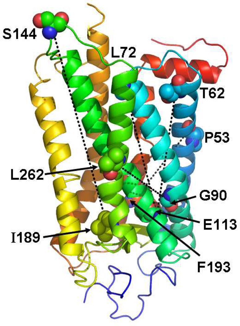
Figure 4.
Rhodopsin structure highlighting the predicted network of communication path from the EC to CP domain. The significant edges representing the predicted network of interactions are highlighted as dashed lines and colored black. The residues that participate in the predicted network of interactions are labeled and rendered as spheres.

