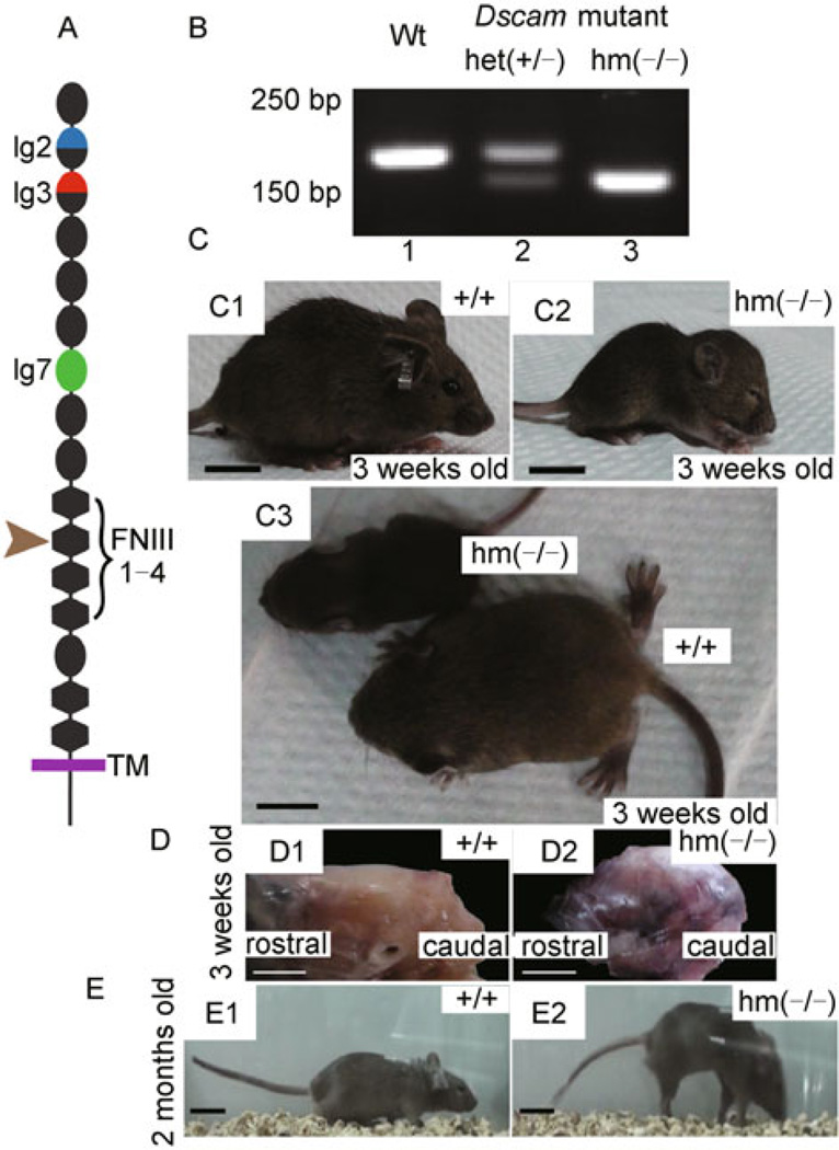
Figure 1. Abnormal body size, head shape and walking posture of homozygous Dscamdel17 mutant mice.
(A) The domain structure of DSCAM. The Dscamdel17 mutation results in a truncated protein at the second FNIII domain. (B) Genotyping of Dscamdel17 mutant mice. PCR with the specific primers led to detection of a 170 bp band in wild type mice (panel 1), a 133 bp band in homozygous Dscamdel17 mutant mice (panel 3), and both bands in heterozygous Dscamdel17 mutant mice (panel 2). (C) Physical characteristics of Dscamdel17 mutant mice. At 3 weeks old, homozygous Dscamdel17 mutant mice show delayed development, exhibiting smaller body size (C3), dome-shaped head, small ears and closed eyes (C2) as compared with their wild type littermates (C1). Moreover, at this age, wild type mice were able to walk whereas homozygous Dscamdel17 mutant mice were not (scale bar: 1 cm) (D) Dome-shaped head of a homozygous Dscamdel17 mutant mouse (D1) as compared with that of the wild type mouse (D2) (scale bar: 0.5 cm) (E) Change in locomotion. The body sizes of homozygous Dscamdel17 mutant mice gradually increased and became similar to that of wild type mice. By 2 months of age, homozygous Dscamdel17 mutant mice (E1) were almost the same as the wild type mice in size (E2). As compared with wild type mice (E2), homozygous Dscamdel17 mutant mice showed abnormal locomotion by walking on their toes with tetanic hind limbs bent back and down-curved tails (E1) (scale bar: 2 cm) [Wt, wild type; het(+/−), heterozygous Dscamdel17 mutant mice; hm(−/−), homozygous Dscamdel17 mutant mice].

