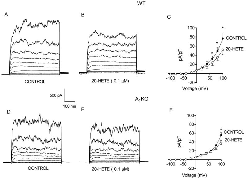
Fig. 2. Effect of 20-HETE on BK current in WT and A1KO aortic myocytes.
Representative current traces are shown under control conditions (A) and with 0.1 μM 20-HETE (B) in a smooth muscle cell from a WT mouse. The voltage template was the same as Fig. 1. (C) Group data (n = 5) show the decrease in the BK current by 20-HETE in smooth muscle cells from WT mice. Representative traces are shown under control conditions (D) and with 0.1 μM 20-HETE (E) for a smooth muscle cell from an A1KO mouse. (F) Group data (n = 5) show the decrease in BK current by 0.1 μM 20-HETE in smooth muscle cells from A1KO mice. *p<0.05 compared to the respective control.

