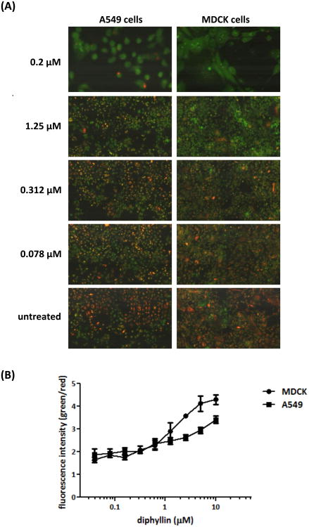
Fig. 2. Dose-dependent inhibition of endosomal acidification caused by diphyllin.
MDCK cells and A549 cells were incubated with bafilomycin A1 (0.2 μM) or various concentrations of diphyllin (0.078, 0.312, 1.25 μM) at 37°C for 20 min. Untreated cells (media only) were used as controls. Acridine orange dye (1 μg/ml) was added to each well and incubated for 10 min. (A) Acidic endosomes in cells were stained red by acridine orange and non-acidic endosomes were stained green. Fluorescence images were obtained on iCys Research Imaging Cytometer. Representative images are shown (magnification: 40×). (B) Fluorescence data was collected from diphyllin-treated wells and the green/red fluorescence ratio was presented. Data in the plot present the mean ± SD out of four replicates.

