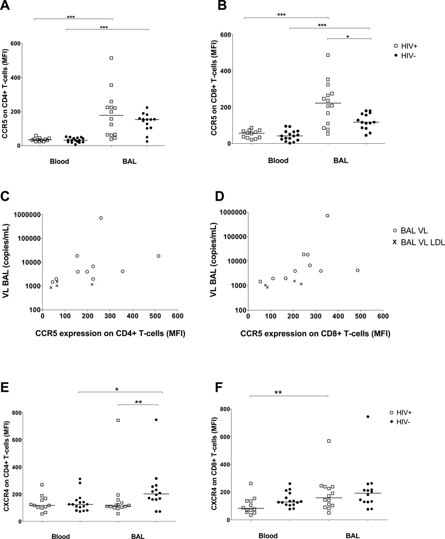
Figure 2.
CCR5 and CXCR4 expression on CD4+ or CD8+ T-cells in blood and bronchoalveolar lavage (BAL). Median fluorescence intensity (MFI) of CCR5 (A,B) and CXCR4 (E,F) receptor expression was measured by flow cytometry on CD4+ (A,E) and CD8+ (B,F) cells from blood and BAL of HIV-1 infected (n=12 in blood, n=14 in BAL, open squares) and HIV-1 uninfected persons (n=16 in blood, n=14 in BAL, solid circles), each sign represents one individual, bars represent medians. Differences between CCR5 expression on CD4+ and CD8+ T-cells from paired blood and BAL samples were calculated by Wilcoxon signed rank test (all p<0.001). The CCR5 expression on CD8+ BAL T-cells was significantly higher in HIV-1 infected compared to HIV-1 uninfected persons (p=0.026, by Mann Whitney U test). Correlation between viral load (VL) in BAL and MFI of CCR5+ on CD4+ (rho=0.706, p=0.005, C) or CD8+ (rho=0.793, p<0.001, D) BAL T-cells of HIV-1 infected participants was assessed by Spearman. Open circles, BAL VL; symbol x, BAL VL lower detection limit (LDL) were set to a value of 19 copies/mL and normalized according to the Urea method. (E) The difference of CXCR4 expression on CD4+ paired blood and BAL T-cells was p=0.049 (Wilcoxon signed rank test). MFI of CXCR4+ CD4+ BALMC was significantly higher in the HIV-1 uninfected control group when compared to HIV-1 infected persons (p=0.009, Mann Whitney U test). BALMC CD8+ T-cells expressed higher levels of CXCR4+ than PBMC in HIV-1 infected persons (p=0.003), differences between the HIV-1 status were assessed by Mann Whitney U test.

