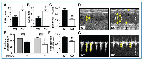
Figure 3. Dilated hypokinetic hearts in cocaine-challenged KATP channel deficient hearts.
Examination by ultrasound revealed dilated hearts in Kir6.2-KO compared to WT after chronic cocaine administration. (A) Left ventricular end-diastolic dimension (LVDd; 3.3 ± 0.2 mm vs. 2.1 ± 0.2 mm; n = 5) and (B) left ventricular end-systolic dimension (LVSd; 2.4 ± 0.2 mm vs. 1.3 ± 0.2 mm; n = 4) were augmented in Kir6.2-KO compared to WT after 20 days of cocaine. (C) The ratio between relative ventricular wall thickness (the sum of interventricular septum thickness, IVST, and posterior wall thickness, PWT) and LVDd was lower in Kir6.2-KO (0.5 ± 0.1; n = 5) versus WT (1 ± 0.1; n = 4) after the cocaine regimen, as shown in representative echocardiographic M-mode recordings (D). (E) Deteriorated cardiac performance following chronic cocaine injections was observed as a significantly reduced fractional shortening from the drug-free baseline in Kir6.2-KO (48.2 ± 4.2% pre- vs. 28.3 ± 4.2% post-cocaine; n = 5 in each group) in contrast to sustained function in WT (47.5 ± 4.2% pre- vs. 35.4 ± 4.7%; n = 4 in each group). (F) Peak ejection velocity appeared lower in Kir6.2-KO (0.49 ± 0.05 m/s; n = 5) versus WT (0.67 ± 0.03 m/s; n = 5), as documented by representative Doppler ultrasound (C). †denotes p < 0.05 when comparing each group to baseline drug-free state. *denotes p < 0.01 when comparing WT versus Kir6.2-KO.

