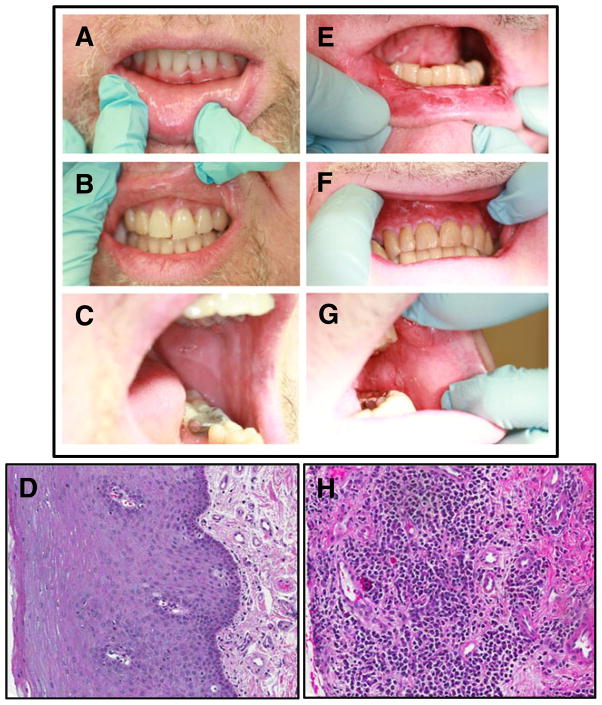
Fig. 1.
Clinical and histological presentation of no oral cGVHD (a–d) versus oral cGVHD (e–h). Clinical presentation of oral cGVHD includes atrophy, erythema, hyperkeratosis, lichenoid reactions, ulcerations, and edema, here seen on the lower lip (e), maxillary anterior gingiva (f), and buccal mucosa (g) of a patient with oral cGVHD, as compared with the normal intraoral mucosa of a patient with no oral cGVHD (ac). H&E staining of the oral mucosa from the above-matched patients demonstrates characteristic GVHD-like changes in the buccal mucosa compared with No oral cGVHD tissue (d), including prevalent lymphocytic infiltrate and apoptotic bodies (h). Buccal biopsy sections are shown in similar orientation at 10x magnification

