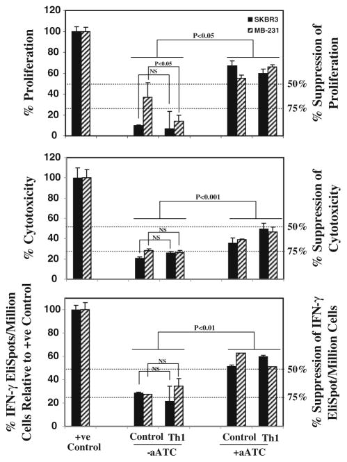
Fig. 4.
Functional properties of MDSC. For analysis of inhibitory ability of MDSCs, CD33+ cells were added at 1:5 (CD33+/aATC) ratio to T-cell proliferation, 1:2 ratio to cytotoxicity and IFN-γ EliSpot assays. Top panel shows the suppressive effect of CD33+ MDSC on anti-CD3-stimulated autologous T-cell proliferation. Proliferation was significantly suppressed by 70–90% in the presence of CD33+ MDSC that was reversed if aATC were present in co-cultures. CD33+ cells isolated from various culture conditions when added to positive controls (Her2Bi-armed ATC-mediated killing of SK-BR-3 for cytotoxicity; SK-BR-3 stimulated Her2Bi-armed ATC for Eli-Spots) showed approximately 75% suppression of cytotoxicity (middle panel) and IFN-γ EliSpots (bottom panel). In the presence of aATC under both control and Th1-enriched culture conditions, suppressive ability of CD33+ cells was significantly attenuated

