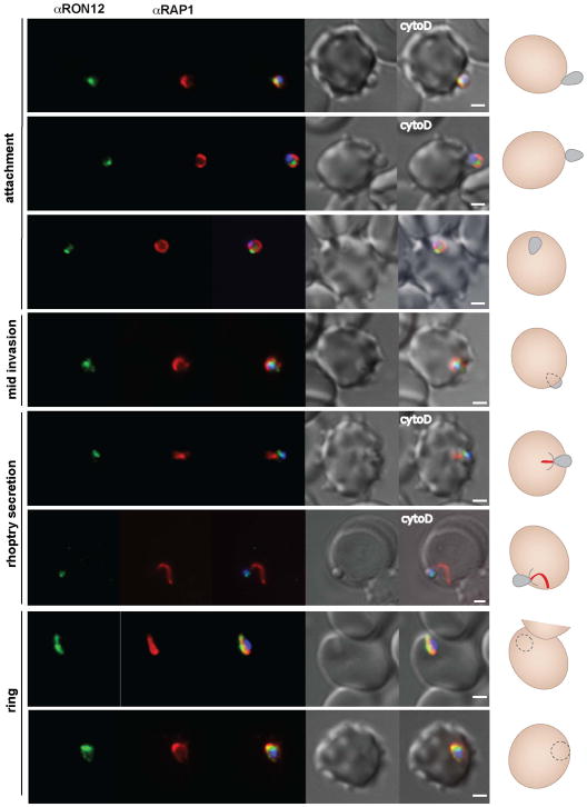
Figure 4. RON12 secretion into the nascent PV during invasion follows that of the rhoptry bulb located RAP1.
Immunofluorescence and DIC images of merozoites fixed during invasion of erythrocytes. From left to right: anti RON12 (green), anti RAP1 mAb 7H8/50 (red), overlay of both with DAPI nuclear stain, DIC image and overlay of all images. Merozoite attachment in the presence of cytochalasin D (cyto D) is indicated. A cartoon schematic is shown on the right of each panel (red line represents RAP1 secreted into the host cell). Size bars equal 1 μm.

