Figure 1.
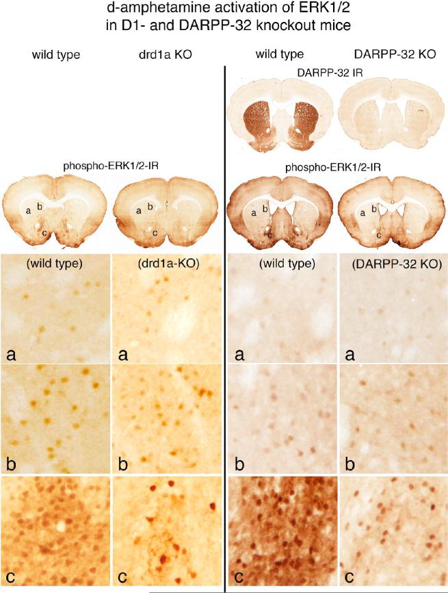
Psychostimulant-induced activation of ERK1/2 in drd1a- and DARPP-32-knockout mice. Effects of d-amphetamine treatment (10mg/kg) are compared between wild type and drd1a knockout (KO) mice and between wild type and DARPP-32 KO mice, in which DARPP-32 immunoreactivity (IR) in the striatum is absent. Activation of ERK1/2 is indicated in coronal brain sections by neurons displaying immunoreactive phosphorylated ERK1/2 (phospho-ERK1/2-IR). In the dorso-lateral striatum (a) and in the dorso-medial striatum (b) the numbers of scattered neurons with d-amphetamine-induced immunoreactive phospho-ERK1/2 is similar in the wild type and drd1a-KO and DARPP-32 KO animals. In the shell of the nucleus accumbens (c), there are numerous d-amphetamine-induced phospho-ERK1/2 immunoreactive neurons in the wild type mice, whereas in both the drd1a-KO and DARPP-32-KO mice the numbers of such neurons is markedly reduced. These data indicate that psychostimulant activation of ERK1/2 mediated by drd1a and DARPP-32 is restricted to the nucleus accumbens.

