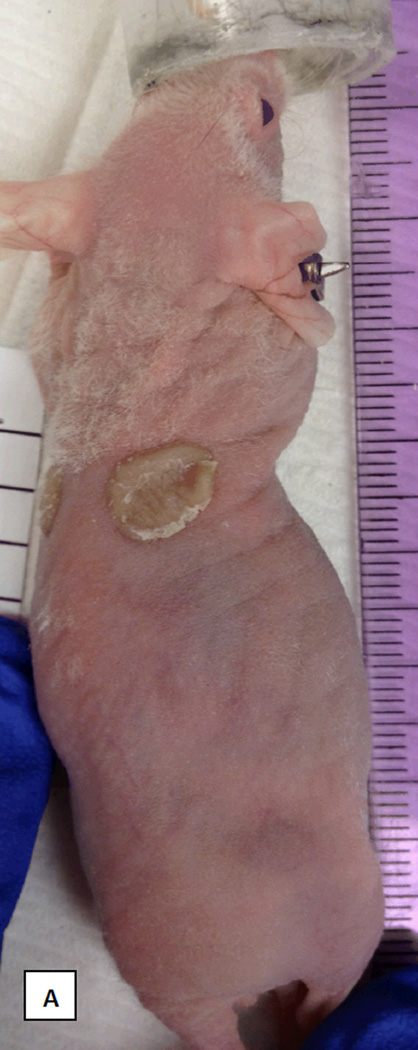
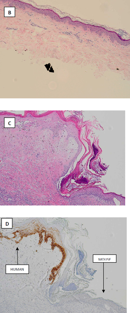
FIGURE 4.
Correlative mouse xenograft model. A: photograph demonstrating successful engraftment of patient skin onto nude mouse at day 31. B: Hematoxyllin and Eosin staining of xenografted tissue prior to transplantation (x4). C: Hematoxyllin and Eosin staining of xenograft after harvest at 87 days post-transplant (x10). D: Staining for human major histocompatability complex-1 (MHC-1) in the harvested graft clearly demonstrating viable epithelial human tissue engrafted adjacent to mouse tissue (x10).


