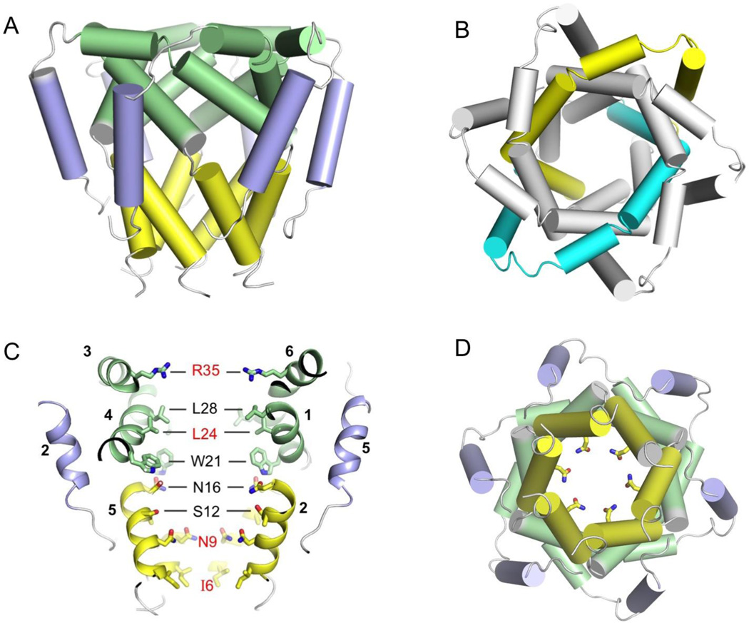
Figure 2. The funnel architecture and pore elements of the p7 channel.
(A) NMR structure of the p7 hexamer. Left panel: cartoon (cylinder) representation illustrating the funnel architecture of the channel. Right panel: global arrangement of H1, H2 and H3 helical segments in the assembled hexamer, showing that the i and i+3 monomers form a symmetric pair in the hexamer.
(B) Pore-lining elements of the p7 channel. Left panel: cutaway view of the channel showing the pore-lining residues, with residues in red being strongly conserved. The numbers next to the helical segments represent the monomers to which the helices belong. Right panel: the view of the N-terminal opening of the channel showing the carboxamide ring formed with Asn9 sidechains.

