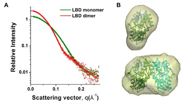
Figure 3.

SAXS data (circles) (A) collected for the GluA2 LBD monomer and the L483Y constitutive dimer. The protein concentration was 0.1 mM in terms of the monomer. Idealized curves (thick lines) (A) were used to generate ab initio models (B) with DAMMIF.16 The monomeric [PDB: 1FTJ, chain A] and dimeric [PDB: 1FTJ, chains A,C] LBD structures4 were fit into surfaces for the respective models.
