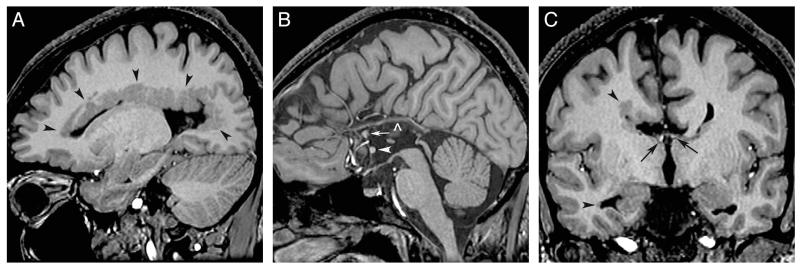
FIG 3.
Heterotopia and commissure anomalies. T1 inversion recovery–weighted images in a 12-year-old boy with dPNH on the right cerebral hemisphere. A, Right parasagittal image shows multiple PNH lining the entire margin of the lateral ventricle. B, Sagittal image demonstrates agenesis of the corpus callosum with a thick anterior commissure (arrowhead) and an anteriorly positioned hippocampal commissure (arrow). A vascular structure is running along of the top of the third ventricle (open arrowhead). C, Coronal image shows moderate white matter volume reduction in the right hemisphere. The fornices are properly located at the roof of the third ventricle (arrows). PNH is seen in the margin of the frontal and temporal horns (arrowheads).

