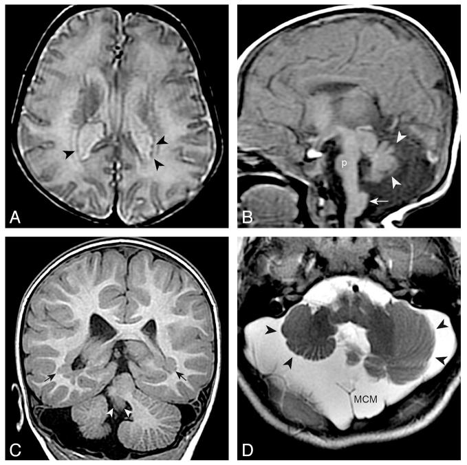
FIG 4.
pPNH and posterior fossa anomalies. A and B, An 11–day–old boy with microcephaly. Small PNH are present in the trigones (black arrowheads in A [axial T2 spin-echo–weighted image]). Other findings include a small pons (p in B [sagittal T1 spin-echo–weighted image]), a hypoplastic and dysmorphic vermis (white arrowheads), and a large inferior cerebellar peduncle (arrow). C and D, A 2-year-old boy with PNH in the trigones (black arrows in C [coronal T1 inversion recovery–weighted image]) and a very small vermis (white arrowheads), dysplastic and small cerebellar hemispheres (black arrowheads), and megacisterna magna (MCM).

