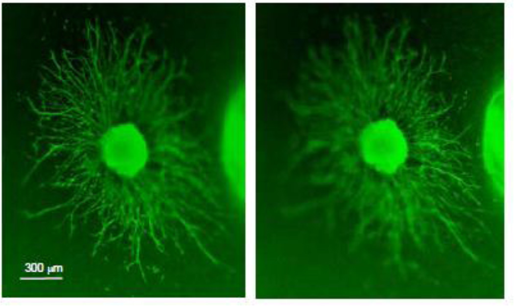
Figure 8. Growth of spiral ganglion neurites in three-dimensional (3-D) plane.
The explant was immunolabeled for neurofilament 200 (FITC). In a collagen gel, neurites grow from a spiral ganglion explant exposed to both neurotrophin-3 and brain-derived neurotrophic factor both in the horizontal and vertical dimensions. The left and right images show two focal planes, in order to illustrate the three-dimensional nature of neurite extension through the gel. The mean length of neurites was shorter than that observed on explants grown on a two-dimensional collagen substrate (Figure 3). The images were obtained on an Olympus IX70 inverted fluorescent microscope.

