Figure 1.
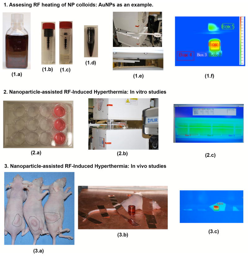
Experimental overview. AuNP heating assessment: As-purchased AuNPs (1.a) are placed in a 50 kDa filter (1.b) and centrifuged down to separate the AuNPs from the filtrate (1.c). This allows for highly concentrated and purified AuNPs to be formed (1.d). The sample is then placed into the RF system using a Teflon sample holder mounted to an adjustable rotary stage (1.e). The AuNPs heating rates, as well as four other control areas, are recorded using an IR camera. In vitro protocols: Hep3B hepatic cancer cells are grown in the front 3-wells of several 12-well cell packs as shown in 2.a (the amount of cell-packs used depends on what the experimentalist wishes to investigate in terms of applied RF power, AuNP concentration, controls, etc.). Each 12-well plate is then subjected to the RF field (2.b). Although not necessary as the optimum RF exposure time has already been determined the media temperature can also be recorded using the IR camera (2.c). In vivo protocols: BALB-C mice beating ectopic hepatic tumors (3.a) were subjected to intra-tumoral injections of AuNPs and exposed to the RF system (3.b) for several minutes. Copper tape was used to ground the mice in order to prevent skin burning. A quartz cuvette filled with AuNPs is also shown next to the mouse to validate RF exposure. The tumor area should have a temperature higher than the rest of the mouse and usually appears red in the IR picture (3.c).

