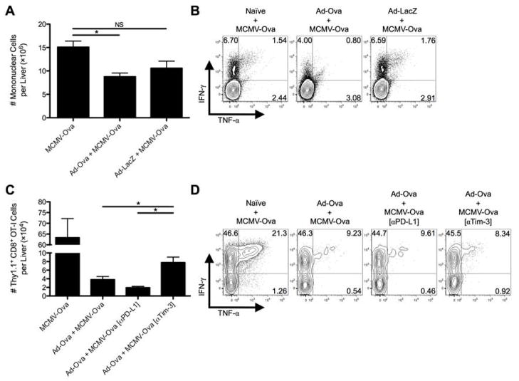
Fig. 5. Tim-3 blockade improves antigen-specific hepatic secondary immune responses to viral infection.
C57BL/6 mice were IV infected with 2.5×10⁷ IU Ad-Ova, 2.5×10⁷ IU Ad-LacZ, or left uninfected. At D7, all 3 experimental groups were IV infected with 1×10⁴ IU MCMV-Ova. (A) The number of live mononuclear cells isolated from the livers of D14 infected animals was enumerated. Trypan blue exclusion was used to assess the number of viable cells. (B) Endogenous CD8+ T cell TNF-α and IFN-γ were quantified at D14 after a 5 hr re-stimulation with 2 μg/mL SIINFEKL peptide (n = 3 per group). (C and D) In a parallel experiment, 5×10⁵ naïve Thy1.1+CD8+ OT-I T cells were transferred at D7. C57BL/6 mice were left untreated or were administered 300 μg anti-PD-L1 Ab or anti-Tim-3 Ab IP at D5 and D6 prior to a D7 MCMV-Ova infection. The number of Thy1.1+CD8+ OT-I T cells and their TNF-α and IFN-γ production was assessed in the livers of infected animals at D14. Cytokine detection was achieved after a 5 hr re-stimulation with 2 μg/mL SIINFEKL peptide (one-way ANOVA/Tukey’s post test; n = 3 per group). Numbers in the scatter plots represent percentages. Mean ± s.e.m.; *P < 0.05.

