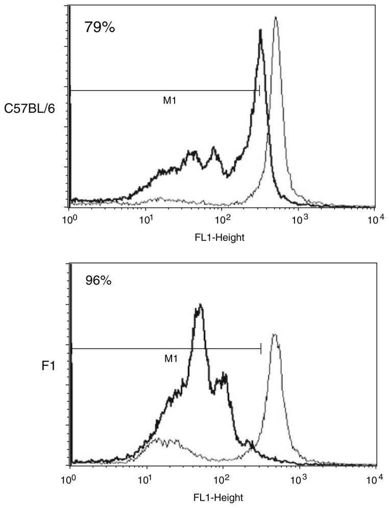
Fig. 2.
Stimulation of antigen-specific T cells by DC in vitro. Transgenic OT1 cells were labelled with CSFE and co-cultured with either SIINFEKL-pulsed (black histogram) or unpulsed (grey histogram) mature C57BL/6 (autologous) or F1 (semi-allogeneic) derived DC in vitro. Flow cytometric analysis of the cells reflects OT1 cell proliferation by dilution of the CFSE dye 72 h later. Results shown from one representative assay from three separate experiments

