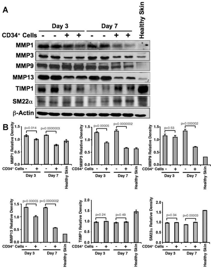
Figure 3. Enhanced fibroblast and myofibroblast cells in response to CD34+ cell therapy.
Immunofluorescence mediated detection of (A) fibroblast (SM22-α), and (B) myofibroblast (alpha smooth muscle actin, α-SMA) cells in the wound bed of animals received CD34+ cells or without cells as a control at various time points of therapy.

