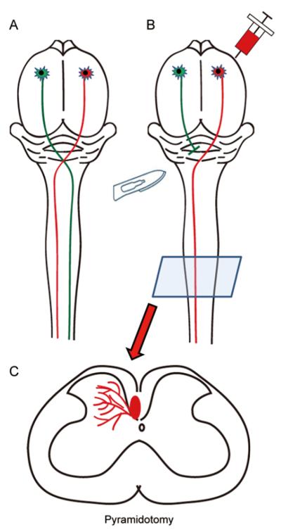
Fig. 3.
Diagram of a pyramidotomy model. Corticospinal axons originate from layer 5 of the motor cortex, decussate at the medullary pyramids and descend the length of the spinal cord without significant bilateral innervation in rodents (A). A pyramidotomy lesions the corticospinal axons at the level of the brainstem before the decussation so that the contralateral spinal cord is virtually devoid of corticospinal innervation (B). Injection of a tracer into the intact tract can be observed at the level of the spinal cord (C) and assessed for axonal growth into the contralateral side.

