Fig. 4.
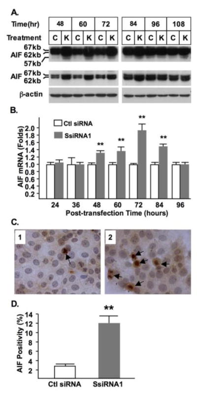
Activation of AIF by syncytin-1 knockdown. (A) BeWo cells were examined for AIF cleavage by Western blot analysis at various post-transfection time points. Positions and sizes of uncleaved and cleaved AIF were indicated. Top panel: long exposure; Mid-panel: short exposure; Bottom panel: β-actin. At 48, 60, 72 and 84 hours post-transfection, increased total AIF protein as well as increased AIF cleavage were detected in the cells transfected with SsiRNA1 (marked as K) compared to cells transfected with control siRNA (marked as C). (B) AIF mRNA levels were determined by real-time PCR. At 48, 60, 72 and 84 hours post-transfection, significantly increased AIF mRNA levels were found in the syncytin-1 knockdown (SsiRNA1) groups compared to control groups (Ctl siRNA) (**, P < 0.01). (C) BeWo cells transfected with control siRNA (panel 1) or syncytin-1-siRNA (panel 2) were examined by immunocytochemistry using the AIF-specific antibody. Nuclei were counterstained with hematoxylin. Increased AIF translocation was observed in a number of cells, which are defined as AIF-positive (marked by arrows). (D) AIF-positive and total cells were counted and the AIF-positivities were compared. A significant increase in the percentage of AIF-positive cells was observed in the syncytin-1 knockdown group (SsiRNA1) compared to the control group (Ctl siRNA) (**, P < 0.01). Note the concentrated staining of nucleolus in some AIF-positive cells.

