Figure 9.
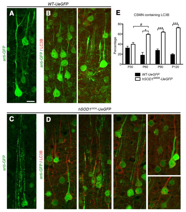
Autophagosome accumulation in the apical dendrites of vulnerable CSMN. A, Representative image of eGFP+ CSMN in the motor cortex of WT-UeGFP mice at P120. B, Autophagosomes are visualized by LC3B expression. C, Representative image of eGFP+ CSMN in the motor cortex of hSOD1G93A-UeGFP mice at P120. The vacuolated apical dendrites of vulnerable CSMN are observed. D, Anti-LC3B immunocytochemistry reveal the presence of autophagosomes, especially along the apical dendrites of eGFP+ CSMN in the motor cortex of hSOD1G93A-UeGFP mice. E, Bar graph representation of average percentage CSMN containing autophagosomes along apical dendrites in WT-eGFP and hSOD1G93A-UeGFP mice at P30, P60, P90, and P120. One-way ANOVA followed by Tukey’s post hoc multiple-comparison test and Student’s t test: #p < 0.05; *p < 0.05; ***p < 0.0001. Error bars indicate SEM. Scale bar, 20 μm.

