
Figure 1. BAK-null mouse embryo fibroblasts (MEFs) are markedly impaired for p53-dependent apoptosis.
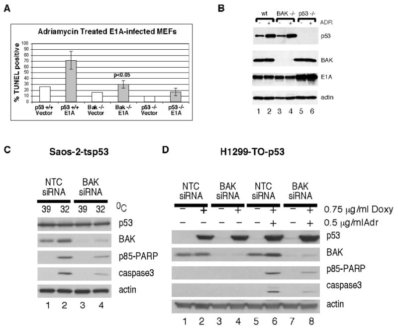
A) TUNEL analysis of apoptosis in MEFs with wt p53 (columns 1 and 2) or from p53-null littermates (columns 5 and 6) or the BAK-null mouse (columns 3 and 4) infected with retrovirus encoding adenovirus E1A (E1A) or viral vector alone (vector) and treated with adriamycin (0.5 ug/mL) for twenty-four hours. Data depicted are the average of three independent experiments, with standard error. B) Western blot analysis of p53, BAK, E1A and β-actin control in lysates isolated from the samples depicted in A, treated or untreated with adriamycin (ADR). C) Saos-2-tsp53 and H1299-Tet-On-p53 cells were transfected with BAK-specific siRNA. Saos-2-tsp53 cells were temperature shifted for 24 hours from 39°C (mutant conformation) to 32°C (wild type conformation) to induce p53. H1299-Tet-On-p53 cells were treated for 3 hours with 0.75 ug/uL of doxycycline, followed by a 20 hour treatment with 0.5 ug/uL adriamycin. p53 expression, actin expression, siRNA mediated knockdown of BAK, and appearance of the caspase cleaved products of PARP (p85 PARP) and caspase 3 were monitored by western blot analysis of whole cell lysates.