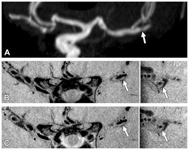
Figure 1.
Grade 0 enhancement of a nonculprit plaque. A, TOF maximum intensity projection MR angiogram of the left middle cerebral artery shows moderate stenosis in the M2 segment (arrow) in a 61-year-old woman. B, Pre- and C, postcontrast 3D VISTA images (left: coronal acquisition) show wall thickening at the corresponding location (arrow). Right: Reconstructions perpendicular to flow direction through the wall thickening show an eccentric atherosclerotic plaque (arrow) with little to no enhancement, similar to that of normal intracranial arterial walls seen elsewhere in this patient.

