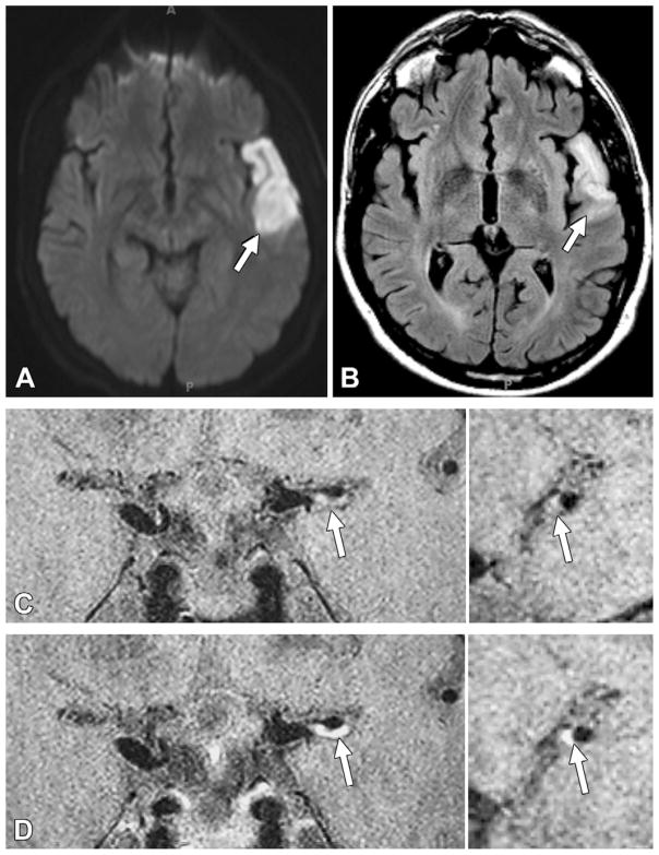
Figure 4.
Acute infarction downstream from a culprit plaque with grade 2 enhancement in a 40-year-old man. A, ■■■ image shows restricted diffusion (arrow) in the left temporal lobe. B, Fluid attenuated inversion-recovery MR image shows corresponding hyperintense signal intensity (arrow). C, Pre- and, D, postcontrast 3D VISTA images (left: coronal acquisition) show eccentric wall thickening, with grade 2 enhancement of the left M1 segment (arrow). Right: Reconstructions perpendicular to flow direction through the wall thickening are also shown.

