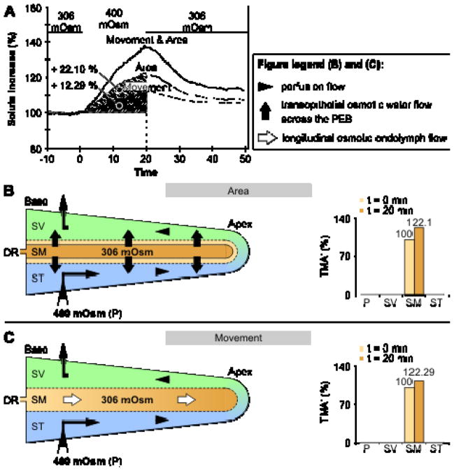
Figure 3.
Mechanisms of endolymphatic volume marker increase during perilymphatic perfusion with hypertonic media in the in vivo experimental study of Salt, DeMott [77]. Hypertonic (400 mOsm (kg·H2O)−1) perfusion of the perilymphatic scalae (scala vestibuli, SV; scala tympani, ST) in the adult guinea pig cochlea induced osmotic volume changes of the endolymph in the scala media (SM). These volume changes were quantified by Salt, DeMott [77] by measuring the concentration change of the ionic volume marker tetramethylammonium (TMA+) after its iontophoretic injection into the endolymph prior to hypertonic perilymphatic perfusion. Salt and DeMott identified two different mechanisms that accounted for the increase in endolymphatic TMA+, i.e., shrinkage of the endolymphatic compartment ((A) and (B), area; TMA+ increased by 22.1%) and apically directed longitudinal flow of TMA+-loaded endolymph ((A) and (B), movement; TMA+ increased by 12.29%). As the epithelial boundary of the cochlear duct is nearly impermeable to TMA+, the induced endolymphatic TMA+ increase can be attributed to a loss of endolymph volume that was proportional to the measured TMA+ increase. DR, ductus reunions; adapted from [77] with permission from the publisher, Elsevier.

