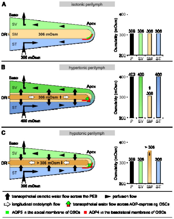
Figure 9.
Hypothetical mechanisms of osmotically induced, AQP4- and AQP5-dependent transepithelial water flow in the cochlear apex and the effect of this flow on longitudinal endolymph flow. (A) In the in vivo experiments by Salt, DeMott [77], perfusion of the scala tympani (ST) and the scala vestibuli (SV) with a solution that was iso-osmotic to the endolymph (306 mOsm (kg·H2O)−1) had no effect on endolymph volume or longitudinal endolymph flow in the scala media (SM). (B) Hyperosmotic (400 mOsm (kg·H2O)−1) perilymphatic perfusion induced shrinkage of the endolymphatic compartment, presumably via osmotically induced transepithelial water outflow along the entire cochlear duct epithelium. Moreover, the longitudinal endolymph flow was into the blind-ending apex of the cochlear duct. This longitudinal flow can be explained by a bulk outflow of water from endolymph across the PEB in the cochlear apex, facilitated by AQP5 in the apical membranes and AQP4 in the basolateral membranes of OSCs in direct contact with the endolymph. (C) When the perilymphatic perfusion solution was changed back to 306 mOsm (kg·H2O)−1, a partial recovery of endolymph volume was measured by Salt, DeMott [77]. This endolymphatic volume increase was potentially induced by transepithelial water inflow along the entire cochlear duct epithelium in response to a reversed perilymphatic-endolymphatic gradient (volume outflow in (C) led to an increase in the levels of endolymphatic osmolytes (> 306 mOsm (kg·H2O)−1)). Consequently, the observed basally directed longitudinal endolymph flow during rehydration of the endolymphatic space [77] may be based on the osmotically induced transepithelial bulk inflow of endolymph through AQP4 and AQP5 in the membranes of OSCs in the apex of the cochlear duct epithelium (DR, ductus reuniens).

