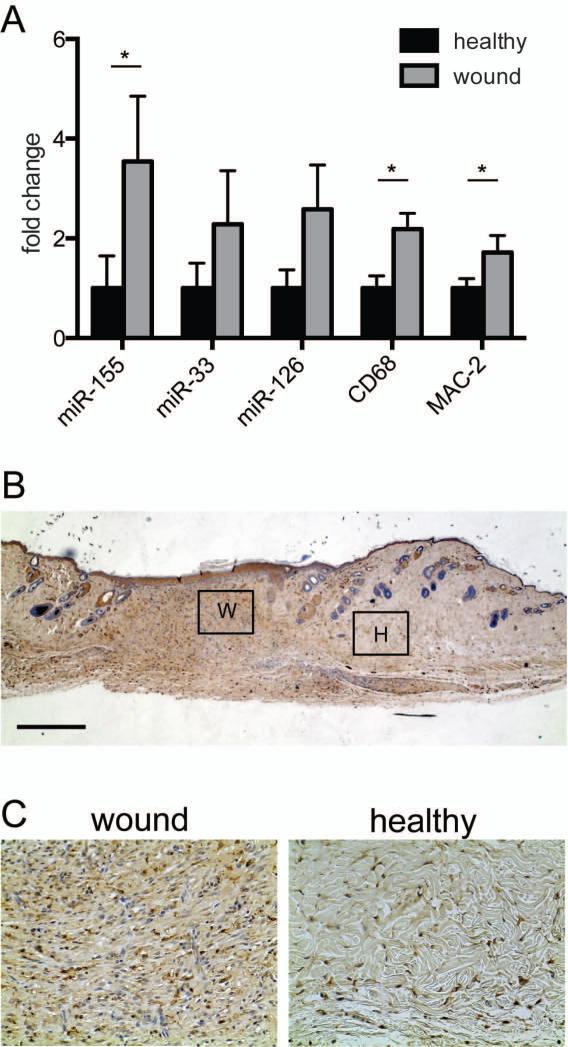
Figure 1. Dermal wounding leads to increased expression of miRs and macrophage markers.
(A) Quantitative analysis of miR-155, miR-33 and miR-126 in wound sections showed that expression levels of these miRNAs are upregulated when compared to healthy skin. qPCR of CD68 and MAC-2 gene expression was used to analyze the influx of macrophages. More macrophages were present in wounded skin when compared to healthy skin. Results are shown as fold difference when compared to mean expression levels of healthy skin using 18S RNA and snord61 as endogenous controls and housekeeping genes for mRNA and miRNA normalization, respectively. Results shown are mean +/- SEM, n=5 animals, *P<0.05 with respect to healthy skin using an ANOVA test followed by a Bonferroni post-hoc test. (B) Representative microscopic image of murine skin (wound and healthy) stained for macrophages with F4/80. Bars indicate 500 μm (C) Areas that are boxed in are zoomed in to show influx of macrophages in wounded (W) and healthy (H) tissue.

