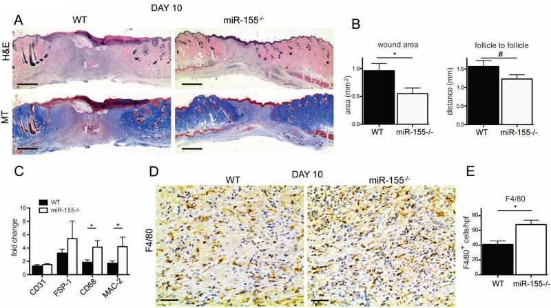
Figure 2. Wounds derived from miR-155–/– mice are smaller 10 days post wounding.
(A) Hematoxylin and eosin (H&E) and Masson's trichome (MT) stained sections demonstrate decreased area of granulation tissue in wounds derived from miR-155–/– animals when compared to WT. Bars indicate 500 μm. A representative microscopic image out of 10 wounds from 10 animals is shown. (B) Left panel, quantification of total wound area demonstrates a significant decrease of wound area in miR-155–/– compared to WT. Right panel, quantification of the distance between bordering healthy skin, depicted by growth of hair follicle, shows a greater distance in WT animals when compared to miR-155–/–. Results shown are mean +/- SEM, n=10 per group, *P<0.05 and #P<0.1 with respect to WT group. (C) Quantitative analysis of the cellular composition of the wound area demonstrated higher expression levels of macrophages makers, CD68 and MAC-1, but not for CD31 and FSP-1, in miR-155-/- group, confirming elevated total numbers of macrophages when compared to WT animals. Results are shown as fold difference when compared to expression levels in healthy skin derived from combining WT and miR-155–/– mice. Results shown are mean +/- SEM, n=10 per group, *P<0.05 with respect to WT group. A representative microscopic image out of 10 stainings form 10 different wounds is shown. (D) Total number of macrophages is elevated in miR-155–/– mice when compared to WT mice. Representative pictures of sections stained with F4/80 in wounded tissue show increased numbers of macrophages (F4/80-positive) in sections derived from miR-155–/– compared to WT. Black bars indicate 50 μm. (E) Quantification of total number of macrophages (F4/80-positive) per high power field (hpf) demonstrates increased numbers of macrophages in miR-155–/– animals.

