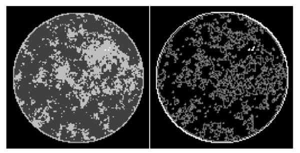
FIGURE 1.

(Left) Discrete phantom modeled after a breast CT application shown in the gray-scale window [0.174 cm−1, 0.253 cm−1]. (Right) Gradient magnitude image (GMI) of the phantom shown in the gray scale window [0.0 cm−1, 0.1 cm−1]. The units of the GMI are also cm−1, because the numerical implementation of ▽ involves only the differences between neighboring pixels without dividing by the physical pixel dimension. The phantom array is composed of 12,892 pixel values, and there are 4,053 non-zero values in the GMI.
