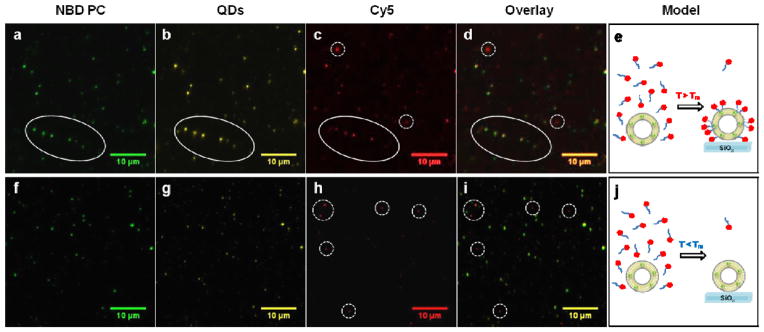
Figure 5. Ligand exchange in lipid membrane embedded CdSe QDs.

Ligand exchange for CdSe QDs encapsulated within DOPC (a–e) and DSPC (f–j) vesicles. Colocalization of fluorescence emission from NBD-PC (a), QDs (b), and Cy5 (c) in individual DOPC vesicles indicates ligand exchange of QDs within fluid lipid vesicles (d and e). (f–j) Shows representative fluorescence microscopy images of QDs embedded within DSPC vesicles. f and g display colocalization, but h show no localization with the QD embedded within the DSPC vesicles, indicating the lack of ligand exchange (i and j). The Cy5 signal in Fig. 5h (dotted circle) is due to the non-specific adsorption of the ligand on the surface of glass, which is also shown in the DOPC-CdSe sample in Fig. 5c (dotted circles).
