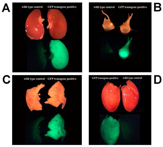
Figure 1.
Example of fluorescence seen in EGFP rat strains. Pictures of organs from SDTg(GFP)2BalRrrc (RRRC:0065) animals (images on right of each panel) and wild type controls (images on left of each panel). Upper images for each panel are under bright light, bottom images for each panel are under fluorescent light. Panel A: kidney; Panel B: eye; Panel C: lung; Panel D: heart.

