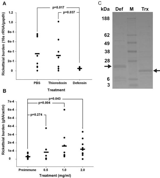
Figure 1. Defensin-2 limits R. montanensis infection in vitro and in vivo.
(A) R. montanensis was incubated with recombinant defensin-2, PBS, or recombinant thioredoxin and then used to infect an L929 monolayer for 24 hours. Rickettsial burden in the infected L929 cells was determined using qRT-PCR. The bar represents the average for each treatment. There are a total of four separate experiments performed in duplicate (two wells per treatment). The number of samples analyzed per treatment are as follows: untreated, n = 8; thioredoxin, n = 8; defensin-2, n = 7. (B) Part-fed D. variabilis ticks were capillary-fed anti-defensin-2 IgG at increasing concentrations followed by an oral R. montanensis challenge. Whole tissue was dissected and rickettsial burden determined using qRT-PCR. Each individual tick is represented by a closed circle. The mean for each treatment is represented by a bar. Values represent the transcript abundance measured in the total number of ticks collected from at least two separate experiments. P-values were determined using the T-Test. Biological replicates are as follows: preimmune, n = 10; 0.5 mg/ml, n = 8; 1 mg/ml, n = 8; 2 mg/ml, 10. (C) Representative Imperial blue stained LDS polyacrylamide gel of recombinant defensin-2 (Def) and thioredoxin (Trx) purification. Defensin-2 migrated to approximately 28 kDa due to the thioredoxin and histidine fusion tags. M, molecular weight marker. Arrows indicate purified recombinant defensin-2 and thioredoxin.

