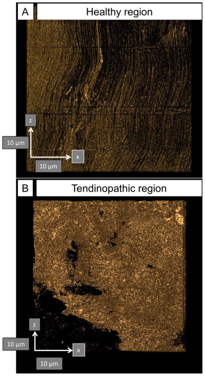
Figure 6. Automated segmentation in IMOD identifies changes in the organization in normal and tendinopathic regions of the same tendon.

(A) Healthy tendon shows near-parallel alignment of collagen fibrils (oriented north-south). (B) Tendinopathic tendon contains disorganized ECM. Arrows show orientation and are 10 μm scale bars.
