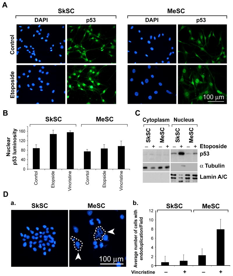
Figure 4. P53 localizes to the nucleus in SkSC and MeSC but the total cellular p53 pool is reduced.
(A) P53 immunofluorescence in SkSC and MeSC treated with etoposide (10 μM, eight hours). Note the predominant nuclear staining for p53 in both cell types, although the p53 signal in MeSC is diminished. (B) The luminosity for the nuclear p53 signal in treated and untreated cells was measured in 10 cells from three random fields and plotted. (C) Western blots of purified nuclear and cytoplasmic fractions from etoposide-treated cells (10 μM, eight hours). As loading controls, blots were stripped and re-probed with γ tubulin (cytoplasm) and lamin A/C (nucleus). (D) Vincristine-treated cells (1 nM, 24 hours) were methanol-fixed and the nuclei stained with DAPI. The circled cells are single cells and the arrows point to endoduplicated nuclei. Cells in ten random fields were counted, and the average number of cells with multiple nuclei (> 3/cell) was plotted.

