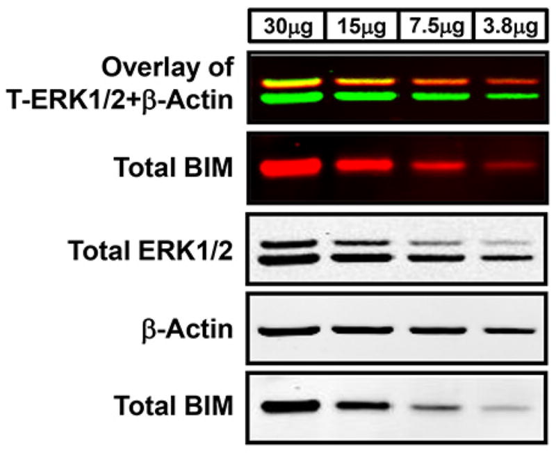
Figure 1. Western blot analysis using two-color infrared fluorescent protein detection.

Lysates of two-fold serial dilutions of WM793 human-derived melanoma protein were separated using a NuPAGE Novex Bis-Tris gel with protein transferred onto PVDF membrane using the iBlot transfer apparatus. Membranes were blocked in Odyssey Blocking Buffer and probed with the following primary antibodies: rabbit anti-total ERK1/2, rabbit anti-total BIM, and mouse anti-β-Actin. Antigen-antibody complexes were detected using fluorescent goat anti-rabbit IRDye 800 (green) or 680 (red) and goat anti-mouse IRDye 680 (red) secondary antibodies, respectively, and visualized with the LI-COR Odyssey Classic Infrared Imaging System. The first Western blot panel is the overlay of the simultaneous detection of total ERK1/2 (800 nm channel-green) and β-Actin (700 nm channel-red) fluorescent signals. The second panel is the single-channel fluorescent detection of total BIM on the same membrane that was separated using a razor blade based on the molecular weights of these target proteins. The lower three Western blot panels are the black and white images of the separated single-channel fluorescent images.
