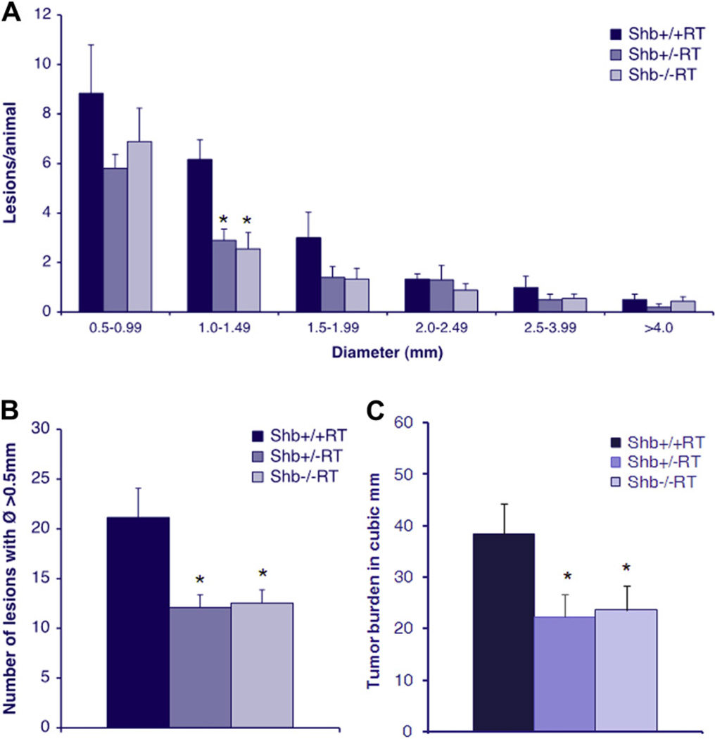
Figure 1.
(A) Lesion size distribution in 12-week Shb+/+RT, Shb+/−RT and Shb−/−RT mice. Number of animals Shb+/+RT n = 6, Shb+/−RT n = 10, Shb−/−RT n = 9, * denotes p-value < 0.05 Student’s t-test when compared wild-type control. (B) Number of lesions with a diameter >0.5 mm in 12-week Shb+/+RT, Shb+/−RT and Shb−/−RT mice. (C) Tumor burden in 12-week Shb+/+RT, Shb+/−RT and Shb−/−RT mice, measured as total volume of lesions with a diameter >0.5 mm. n = 16–24.

