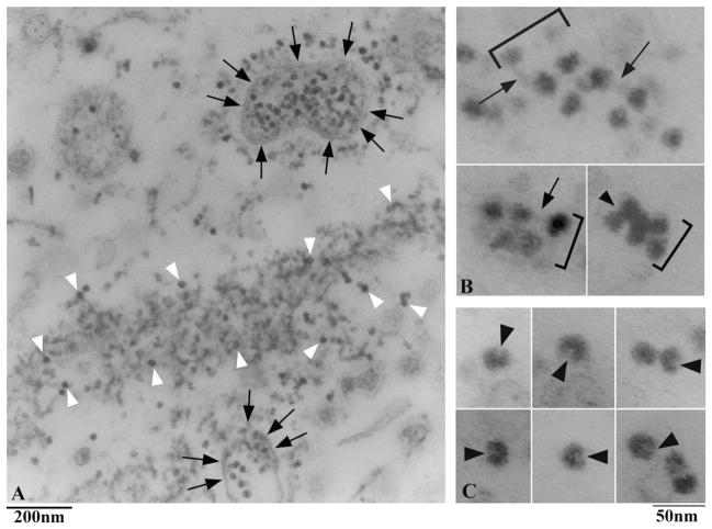
Fig. 7. Thin sections of the gradient pellet.
A. Low magnification view. IFT particles appear as electrondense particles, some of which are here indicated by white arrowheads. Particles often associate to form large aggregates (as the one containing the particles indicated by the white arrowheads), or are associated with membrane vesicles (indicated by the black arrows). B. Higher magnification views of short, double row arrays of particles, which are indicated by brackets. The arrows indicate the interconnecting links between adjacent particles. The arrowhead indicate a double row array of particles showing a more compact organization. C. In some sections, particles appear to possess a central cleft (arrows).

