Figure 12.
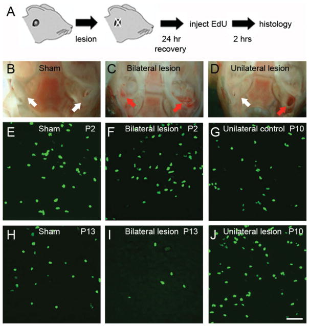
Bilateral ear lesions affect cell proliferation in the rat MNTB. A: Experiment design. Ear lesions were performed as described in Materials and Methods and proliferating cells were labeled with EdU histochemistry after a recovery period of 24 hours. B–D: Exemplar images show the extent of damage in the inner ear of sham (B), bilateral lesion (C), and unilateral lesion animals (D). White arrows indicate intact inner ear. Red arrows indicate lesioned inner ear. E–J: Exemplar images of EdU stained brainstem slices from sham (E,H), bilateral lesion (F,I), and unilateral lesion experiments (G,J). The unlesioned side was used as a control for unilateral lesions. Scale bar = 50 μm in J (applies to E–I).

