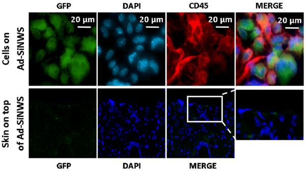
Figure 6.

Ex vivo fluorescence imaging of cells located on the Ad-SiNWS and skin tissue on top of the Ad-SiNWS after in vivo NSMD of pEGFP. The micrographs indicated that the immune cells (CD45 positive) were recruited onto the Ad-SiNWS and were transfected with pEGFP⊂SNPs. The cells on the skin tissue section, however, showed no significant fluorescence signals, suggesting that the skin cells on top of Ad-SiNWS were not transfected with pEGFP in the process of the in vivo NSMD study.
