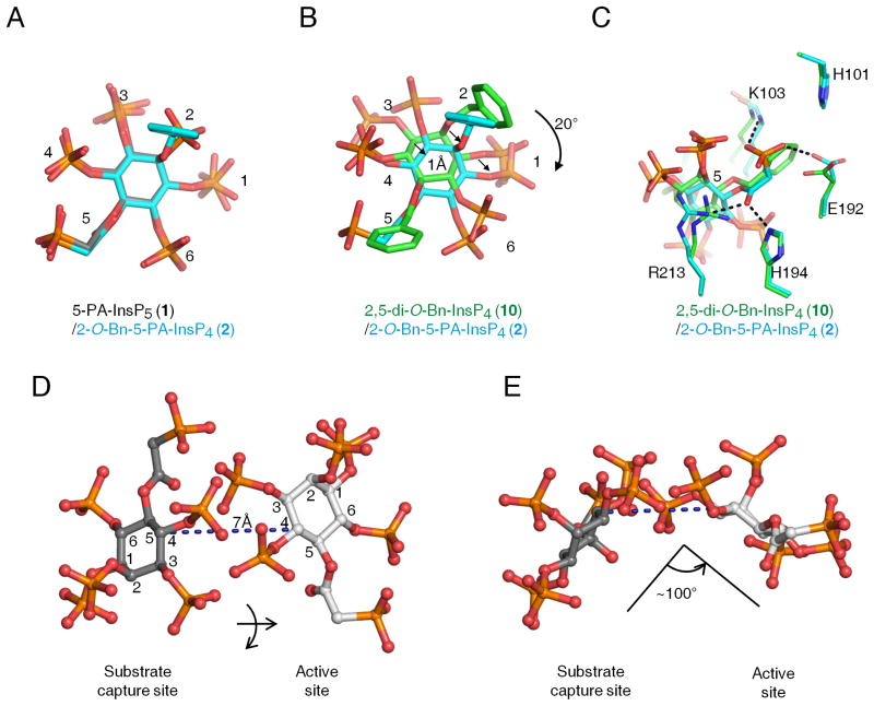
Figure 6. Relative orientations of inositol phosphate analogues in the second ligand binding site of PPIP5K2KD.
Data for ligand orientation in the second binding site were obtained as described in the Figure 5 legend. Analogs are depicted as stick models. Atoms are colored blue for nitrogen, red for oxygen and orange for phosphorus. Panel A superimposes 5-PA-InsP5 (1) (carbons colored gray) and 2-O-Bn-5-PA-InsP4 (2) (carbons in cyan). Panel B and C superimpose 2,5-di-O-Bn-InsP4 (10) (carbons colored green) and 2-O-Bn-5-PA-InsP4 (2) (carbons in cyan). Arrows in panel B emphasize spatial separation, and panel C includes details of ligand interactions with neighboring amino-acid residues. Panels D and E illustrate that transfer of 5-PA-InsP5 (1) between the two sites involves a lateral migration of 7 A, a ring flip, and a rotation of about 100. Atoms are colored blue for nitrogen, red for oxygen, orange for phosphorus, and gray for carbon. The inositol ring is numbered.

