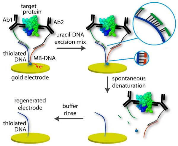
Figure 1.
Cycle for gentle, enzymatic regeneration of the SAM-coated electrode surface. After measurement (upper left), electrode was incubated in uracil-DNA excision mixture (upper right). Cleavage of the DNA backbone occurred at strategically placed uracils. Resulting shorter DNA sections spontaneously denatured (lower right) and were washed away in buffer (lower left), leaving the SAM of thiolated DNA that was ready for probe loading and another measurement.

