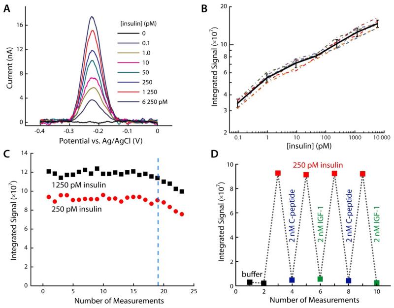
Figure 4.
Antibody based ECPA for insulin quantitation. (A) SWV scans for insulin quantitation in the fM to nM range. Seven different sensors responded proportionally, suggesting high method precision. (B) Insulin calibration curves with seven different sensors (dotted lines), with the average trace (black line) shown; error bars represent standard deviations. The insulin LOD was found to be 10 fM. (C) Two independent sensors were cycled through the reusable methodology during measurement of varying insulin levels (red and black points). Again, up to 19 uses were permitted without significant loss of signal. (D) High target specificity was confirmed by challenging the sensor with a protein of similar structure (IGF-1) and a co-secreted hormone (C-peptide). Only insulin generated measurable responses.

