Fig. 1.

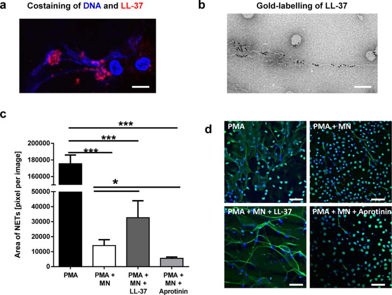
LL-37 protects NETs against degradation by S. aureus nuclease. (a and b) Confocal immunofluorescence (a) and electron micrograph (b) of structures e.g. NETs and nanoparticles released by PMA-activated neutrophils which are associated with a high amount of LL-37. Bar 8 μm (a) and 100 nm (b). (c) Human blood-derived neutrophils were stimulated with 25nM PMA for 4 hours to induce 100% NET formation. Aprotinin (40 μg/ml) was used to block formation of endogenous active LL-37. Then, the NETs were treated with 0.01 U/ml S. aureus nuclease (micrococcal nuclease, MN) in the presence or absence of externally added LL-37 (5 μM). (d) Representative fluorescent micrographs displaying the results of the column bar graph in (c). The bars indicate 50 μm. NETs were visualised using an Alexa 488-labelled antibody against H2A-H2B-DNA complexes (green) in combination with DAPI to stain the nuclei in blue. The area covered with NETs was quantified using Image J. The graph represents the mean ± SEM of minimum 18 images derived from 3 independent experiments. * p < 0.05, *** p < 0.001 by t-test.
