Fig. 3.

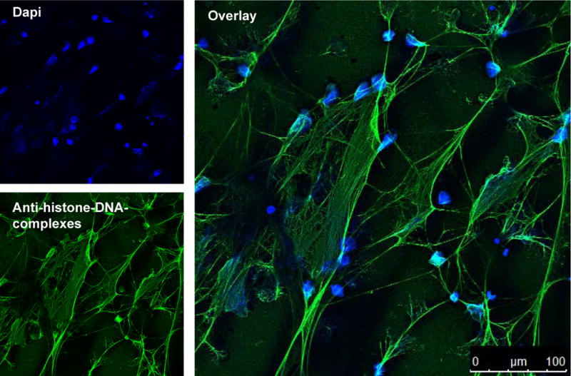
DAPI and immunostaining of NETs. Confocal immunofluorescent micrograph of NETs. NETs were visualised using an Alexa 488-labelled antibody against H2A-H2B-DNA complexes (green) and with DAPI to stain the extra- and intracellular DNA in blue.

DAPI and immunostaining of NETs. Confocal immunofluorescent micrograph of NETs. NETs were visualised using an Alexa 488-labelled antibody against H2A-H2B-DNA complexes (green) and with DAPI to stain the extra- and intracellular DNA in blue.