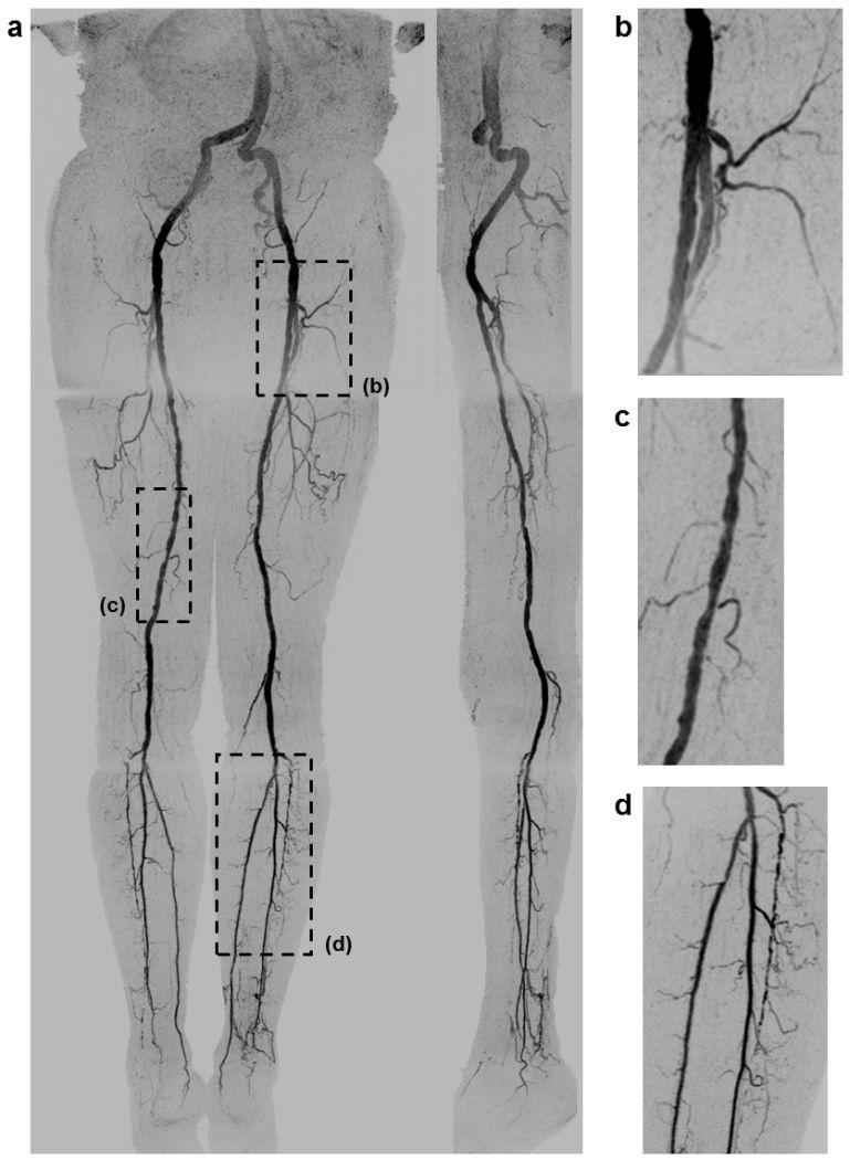
FIGURE 3.

(a) Coronal and sagittal (left leg) MIPs of a healthy 81 year old male volunteer (Subject 4) generated using the final abdomen-pelvis and thigh station time frames and the third calf-foot time frame. The dwell time at the thigh station was only 5.0 sec. Targeted coronal MIPs of the boxed regions in (a) are shown in (b–d), highlighting the arterial detail. Although this subject was recruited as a healthy volunteer, multiple low grade stenoses are seen in the thigh and calf arteries, and the anterior tibial arteries are partially occluded. The contrast bolus was imaged at four abdomen-pelvis and two thigh station time frames, and venous contamination was avoided at the calf-foot station. Reproduced with permission from Ref. (17). Also see Supplemental Figure 1 and Movie 1.
