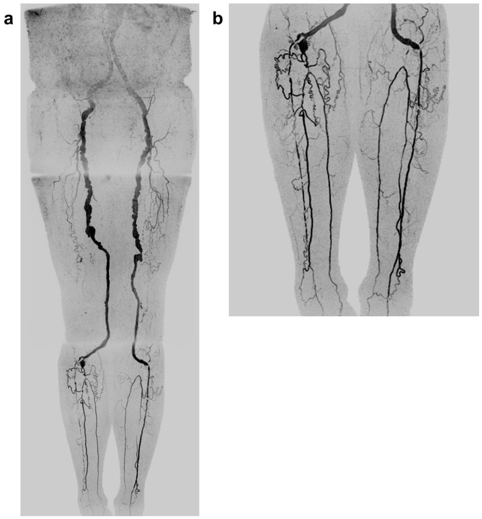
FIGURE 4.

Coronal MIPs of the (a) extended FOV and (b) calf-foot station of an 81 year old male patient (Subject 10) with slow contrast bolus progression. The transit times at the abdomen-pelvis and thigh proximal stations were 10.0 and 12.5 sec, respectively. The extended FOV angiogram was generated using the last abdomen-pelvis and thigh time frames and the second calf-foot frame. The calf-foot MIP shows the third time frame, which has excellent arterial contrast and detail. The calf vasculature has extensive disease and complex flow, including occlusion of both tibioperoneal trunks and filling of the posterior tibial and peroneal arteries via communicating vessels. This study was scored substantially better than the CTA due to the CTA outstripping the bolus on the first run and the second run having suboptimal contrast and anatomical coverage. Also see Supplemental Movie 2.
